Research
Ca2+ imaging and optogenetic stimulation of brain activities by two-photon holographis microscope
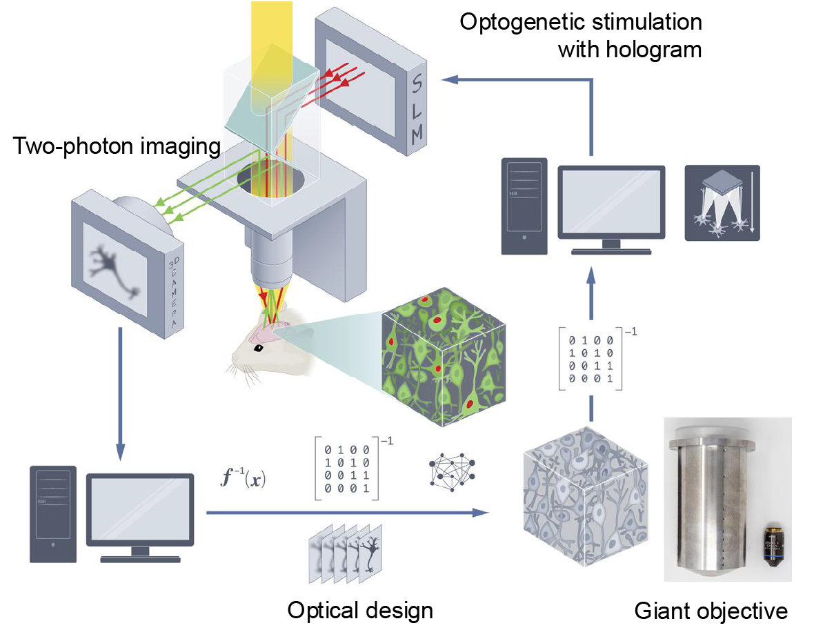
(a) Two-photon holographic microscope
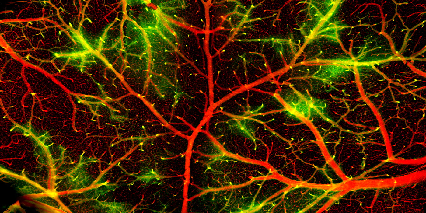
(b) Two-photon Imaging of cerebral neurons and vasculature
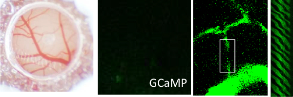
(c) Optogenetic stimulation of cerebral neurons by using hologram
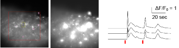
The brain is one of the final frontiers. The mechanisms by which cell activity in the brain gives rise to human consciousness, emotion, thought, etc. have yet to be elucidated. This is because it is difficult to visualize and manipulate the vast cell activity in the brain, and therefore we have not yet obtained sufficient data to understand the brain. It is not easy to visualize and manipulate the inside of the brain using a microscope. This is because the refractive index of the cell membrane and water are very different, so light scatters and the image becomes distorted. To solve this problem, two-photon microscopes were developed that use near-infrared light, which is less prone to scattering, to enable fluorescence imaging of brain tissue to a depth of around 1 mm from the surface. The two-photon holographic microscope is a next-generation two-photon microscope developed at the Kobe University Center for Next-Generation Optical Scattering Imaging Science (OaSIS) (Fig. a), and in addition to fluorescence imaging of neural activity and blood flow in the brain (Fig. b), it is possible to optogenetically stimulate cells using three-dimensional patterns of light called holograms (Figure c). At OaSIS, we are currently conducting research aimed at visualizing and manipulating cells throughout the entire mouse brain by combining a large objective lens and light design-based scattering correction. Our laboratory is participating in this research from a biological approach, and together with experts in optics and information science, we are trying to overcome the limitations of “visualizing and manipulating cell activity in the brain”. We are also trying to find clues to understanding brain function and pathology from brain cell activity that has been revealed for the first time through the development of new microscope systems.
AQP4-dependent astrocytic regulation of neuromodulators and psychiatric disorder
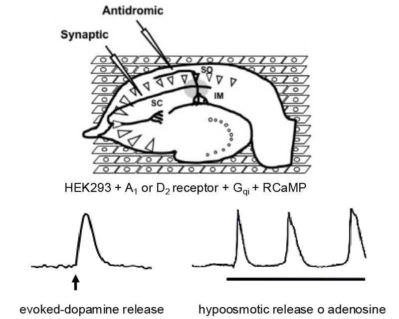
(a) Measurement of meuromodulator release
(b) The up-regulation of AQP4-dependent adenosine release suppresses dopamine neurotransmission during depressions.
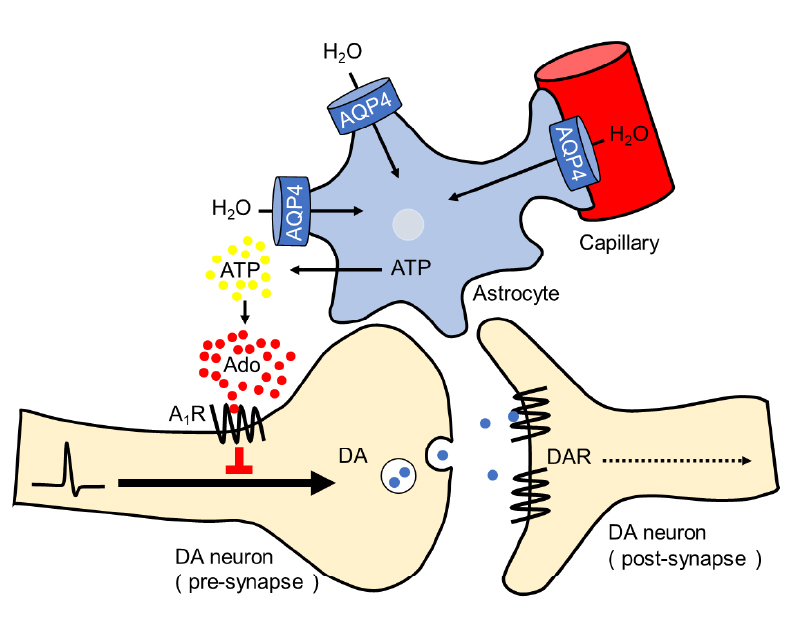
Brain activity is regulated by neuromodulators such as adenosine, dopamine, serotonin, and noradrenaline. Increases or decreases in these modulators are thought to underlie psychiatric disorders such as depression and schizophrenia. Therefore, we analyzed neuromodulators during mouse depression-like behavior, which works as a model for understanding human motivation. Our research has revealed that the water channel (AQP4) in astrocytes, a type of glial cell, regulates the levels of adenosine and dopamine, and that abnormalities in this process cause depressive-like behavior. In this research, we created sensor cells that increase fluoresce in response to adenosine or dopamine using genetic engineering, placed brain slices on top of these cells, and measured dopamine release in response to electrical stimulation and adenosine release in response to hypoosmolality (Figure a). Cocaine, a type of stimulant drug, can cause depression as a withdrawal symptom, so we made brain slices from mice that had been administered cocaine and used them in the measurements. We found that, in association with depressive-like behavior, AQP4-dependent adenosine release increased, and that this adenosine suppressed dopamine neurotransmission (Figure b). In fact, AQP4-deficient mice do not show depressive-like behavior induced by cocaine. Currently, we are conducting research using two-photon holographic microscopy, focusing on points such as “how does cocaine administration affect brain activity?” and “does this effect change in AQP4-deficient mice?”.
Reactive astrocyte and wound healing after closed-head injury
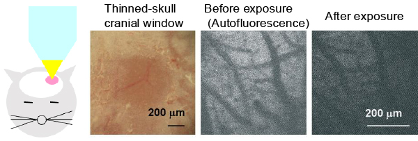
(a) Photo-injury mouse
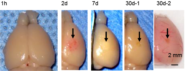
(b) Wound healing
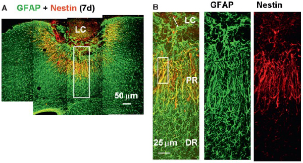
(c) Nestin-expressing reactive astrocyte (NRA)
(d) NRA and woudn healing
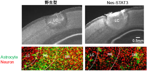
Neurons and glial cells are produced from neural stem cells (NSCs) during development. Recently, NSCs have been discovered in adults after the end of development, and it has become thought that the brain has the capability to regenerate. We have developed a focal brain injury model called photo-injury mice, and we are studying the mechanism of brain regeneration. Photo-injury, which was developed by Morita at the University of New Mexico Department of Neurosurgery, induces focal cerebral degeneration by intense light irradiation through thinned-skull cranial window (Figure a) . In this model, there are no abnormalities in the brain tissue immediately after light irradiation, but after one day, bleeding occurs, and after one month, part of the brain tissue is lost and a brain contusion is formed (Figure b). This wound healing is very similar to the head contusions seen in humans in traffic accidents and sports injuries. In addition, in the photo-injury mice, there is a significant regeneration of brain tissue, and reactive astrocytes that express a neural stem cell marker called nestin (nestin-expressing reactive astrocyte, NRA) (Fig. c) accumulate in the regenerated region. Furthermore, when the STAT3 gene, which is essential for the conversion of normal astrocytes to reactive astrocytes, is disrupted, tissue regeneration does not progress, and the damage remains large (Fig. d), so NRA plays an essential role in brain tissue regeneration. We are currently using two-photon holographic microscopy to study questions such as “How do NRAs regenerate brain tissue?” and “How does wound healing affect brain activity?”. By studying the process by which brain functions and neural circuits lost due to injury are reconstructed, we hope to be able to reveal which neural circuits are responsible for brain functions such as sensation and movement.
BBoad impairment of cerebrospinal fluid circulation following focal brain injury
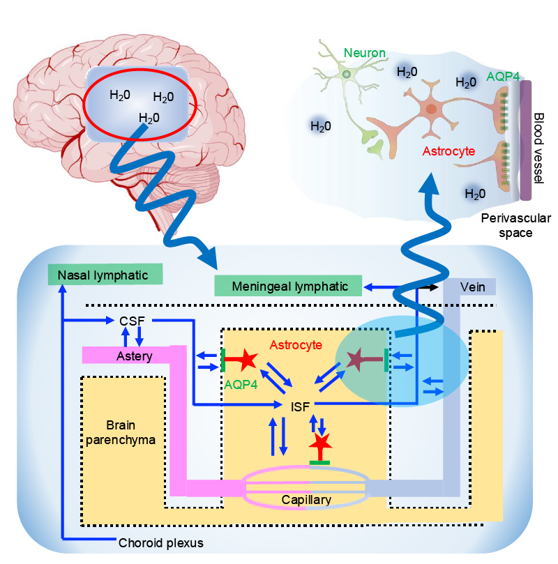
(a) Cerebrospinal fluid (CSF) and AQP4
(b) Cerebral blood flow (CBF) and CSF after photo-injury
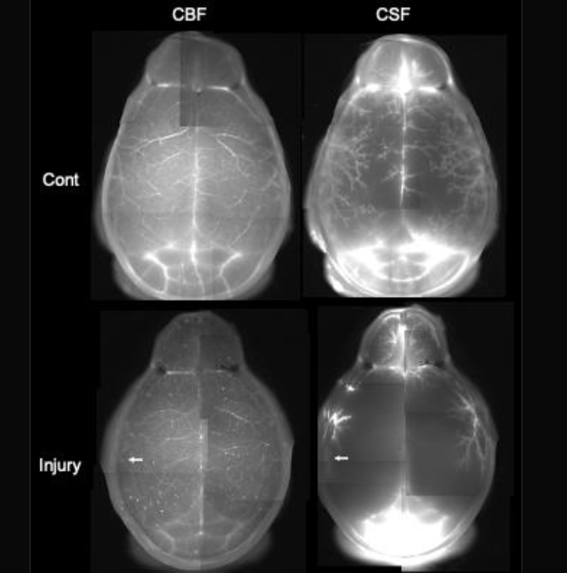
Our bodies have two water circulations: the vascular system and the lymphatic system. However, the brain does not have a lymphatic system, and instead has developed its own water circulation system, which is made up of cerebrospinal fluid (CSF) and the water channels (AQP4) of astrocytes. Although it is still not fully understood how CSF flows, it is proposed that that CSF flows through cerebral perivascular spaces, circulating inside and outside the brain tissue (Figure a). In addition, AQP4 is highly localized at the boundary between astrocytes and blood vessels, and it is known that the circulation of the Glymphatic system is significantly reduced in AQP4-deficient mice. For this reason, it is thought that the above-mentioned AQP4-dependent adenosine release is activated in the process of water flowing into the brain from blood vessels. In order to investigate the CSF circulation in photo-injury mice, we injected different fluorescent dyes into the cerebral blood flow (CBF) and CSF, and double-labeled them (Figure b). As a result, we found that in photo-injury mice (arrow), the CSF, which was distributed along the blood vessels on the surface of the brain in normal mice (Cont), was reduced throughout the brain. When part of the brain is damaged, it is thought that the activation of astrocytes and other factors causes a decrease in water influx via AQP4, and that this worsens the circulation of CSF throughout the brain. These results may be related to the fact that focal brain injuries caused by traffic accidents or sports accidents, can lead to a general decline in brain function and the development of mental illnesses such as depression. We are investigating the possibility that focal brain injury modulates CSF circulation and the aforementioned AQP4-dependent adenosine release throughout the brain, and has various effects on brain function, using two-photon holographic microscopy. If this reveals the reality that brain activity is linked to water circulation, it is expected to provide a new perspective for elucidating brain function and disease.